Rapid ECG Interpretation resources, like those by M. Gabriel Khan, are vital for healthcare professionals. Mastering quick analysis, often found in PDF guides, improves patient outcomes significantly.
The Importance of Quick and Accurate ECG Analysis
Rapid and precise ECG analysis is paramount in clinical settings, directly impacting patient care and survival rates. Delays in identifying critical abnormalities – such as those detailed in resources like “Rapid Interpretation of ECG” PDFs – can lead to worsened outcomes. Accurate interpretation facilitates timely interventions for conditions like myocardial infarction and life-threatening arrhythmias.

The ability to swiftly decipher ECG patterns, gathered from diverse patient cases and readily available in educational materials, empowers healthcare professionals to make informed decisions. Understanding the fundamentals, often emphasized in concise textbooks and online guides, allows for efficient triage and appropriate treatment strategies. A solid grasp of ECG principles, reinforced through practice with case studies (often found in PDF format), is therefore non-negotiable for effective emergency response and patient management.
Target Audience: Who Benefits from Rapid Interpretation Skills?
A broad spectrum of healthcare professionals benefits from mastering rapid ECG interpretation. This includes physicians (emergency medicine, cardiology, internal medicine), nurses, paramedics, and even physician assistants. Resources like the “Rapid Interpretation of ECG” textbook, often available as a PDF, cater to varying levels of expertise.
Specifically, those frequently encountering patients with cardiac complaints – such as emergency room staff and critical care teams – find these skills invaluable. Even clinicians outside of cardiology can utilize basic ECG interpretation to quickly assess patients and determine the need for further evaluation. Furthermore, students in healthcare programs benefit immensely from early exposure to ECG analysis, utilizing online tutorials and case studies (often in PDF form) to build a strong foundation. Ultimately, anyone involved in patient assessment can improve care with this knowledge.

Basic ECG Principles
Understanding waveforms (P, QRS, T), paper speed (25mm/s), and amplitude are foundational. PDF guides like “Rapid Interpretation of ECG” simplify these core concepts.
Understanding the ECG Waveforms (P, QRS, T)
Deciphering ECG waveforms – P, QRS, and T – is central to rapid interpretation. The P wave represents atrial depolarization, initiating the heartbeat. The QRS complex signifies ventricular depolarization, the largest electrical impulse, and dictates ventricular contraction. Finally, the T wave illustrates ventricular repolarization, preparing the ventricles for the next cycle.
Accurate identification of these waveforms is crucial. Resources like M. Gabriel Khan’s “Rapid Interpretation of ECG” (often available as a PDF) provide detailed illustrations and explanations. Understanding their morphology – shape, size, and duration – allows for quick assessment of cardiac function. Abnormalities in any waveform can indicate underlying pathologies, from atrial enlargement to ventricular ischemia. Mastering these basics, readily accessible in focused PDF guides, forms the cornerstone of efficient ECG analysis.
Furthermore, recognizing the timing and relationship between these waves is paramount. A systematic approach, reinforced by practice with ECG databases (available in PDF format), builds confidence and speed.
ECG Paper and Calibration: Speed and Amplitude
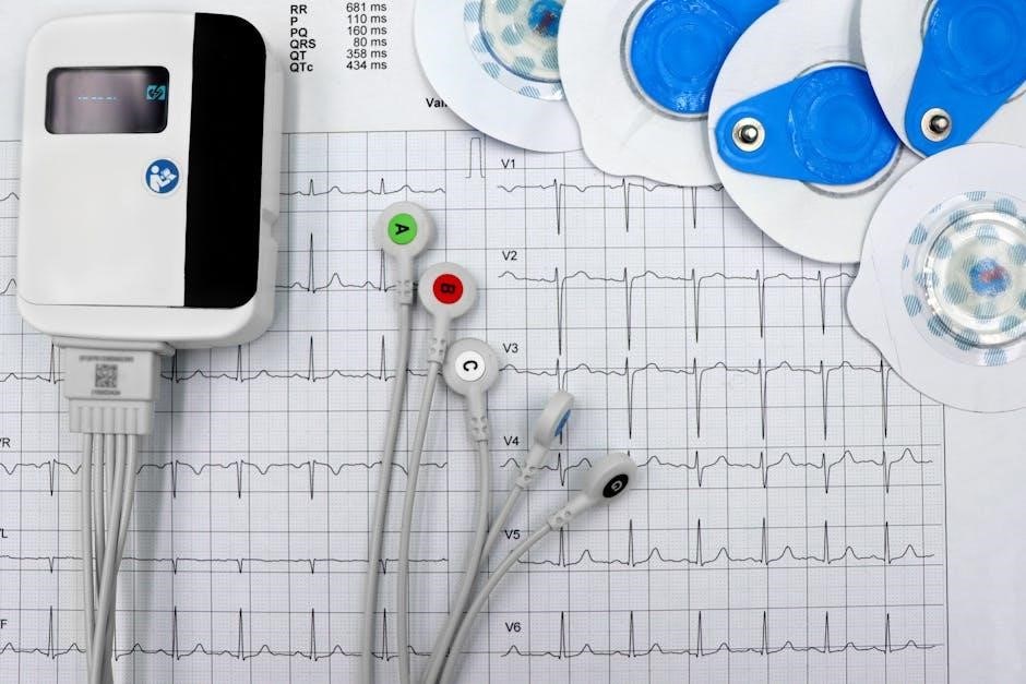
Standard ECG paper moves at a speed of 25 mm/second. This consistent speed allows for accurate time measurements, crucial for rate and rhythm determination. Each small square on the paper represents 0.04 seconds, a fundamental element in rapid interpretation. Understanding this timing is often emphasized in introductory PDF guides.
Amplitude, or voltage, is measured vertically. Each small square represents 0.1 mV. Calibration ensures accurate voltage readings; a standardized 1 mV signal is used for verification. Incorrect calibration can lead to misdiagnosis, highlighting the importance of checking this before analysis. Many “Rapid Interpretation of ECG” resources, including downloadable PDF versions, detail calibration procedures.
Resources, like those found in Harvard-affiliated hospital files (often compiled into PDF collections), demonstrate the impact of paper speed and amplitude on waveform appearance. Consistent calibration and awareness of standard paper speed are foundational skills for any ECG interpreter.
Lead Placement: A Brief Overview of Standard Leads
A 12-lead ECG provides a comprehensive view of the heart’s electrical activity. Standard leads include limb leads (I, II, III, aVR, aVL, aVF) and precordial leads (V1-V6). Correct lead placement is paramount for accurate interpretation; misplacement can mimic pathology. Many introductory PDF guides emphasize proper technique.
Limb leads assess electrical activity in the frontal plane, while precordial leads view the heart from a horizontal plane. Understanding which lead “sees” which part of the heart is key to localizing abnormalities. Resources, like those detailing ECG patterns from patient records (often available as PDFs), demonstrate how lead placement impacts waveform morphology.

“Rapid Interpretation of ECG” materials, including downloadable PDFs, often include diagrams illustrating correct lead placement. Mastering lead recognition and understanding their respective views is a foundational step in efficient ECG analysis, enabling quick and accurate diagnoses.
Systematic Approach to ECG Interpretation
A structured approach, detailed in PDF guides like “Rapid Interpretation of ECG”, is crucial. Focus on rate, rhythm, P waves, QRS, and intervals for efficient analysis.
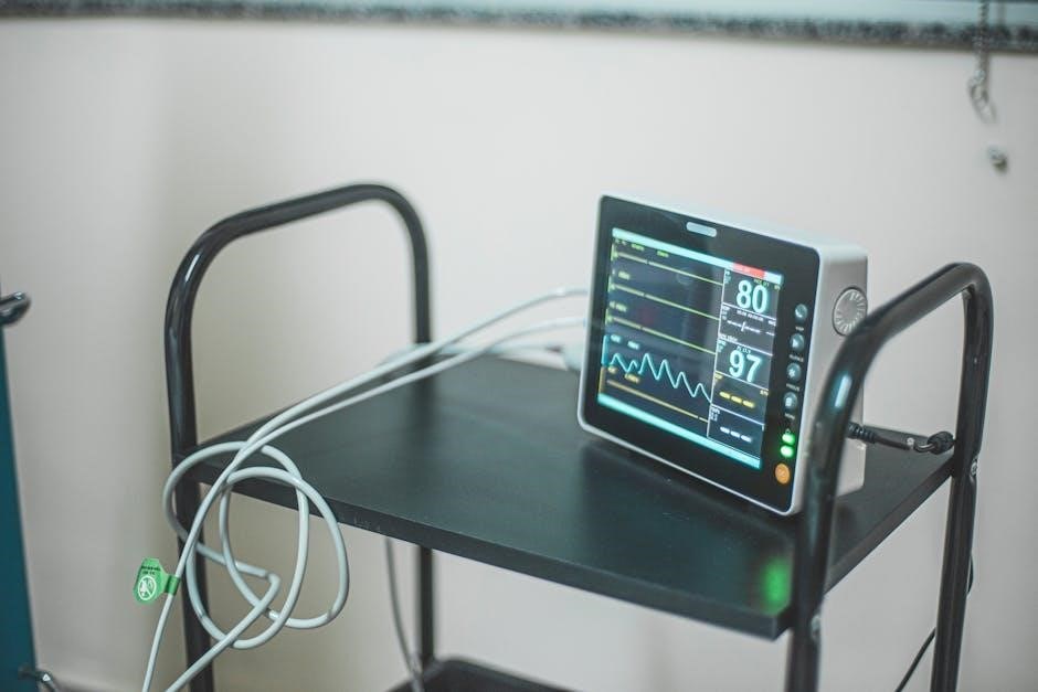
Step 1: Rate Determination – Calculating Heart Rate
Accurate rate determination is the foundational first step in ECG interpretation, frequently detailed in resources like the “Rapid Interpretation of ECG” PDF guides. With a standard ECG paper speed of 25 mm/s, each small square represents 0.04 seconds. Several methods exist for quick calculation.
For regular rhythms, the ‘300, 150, 100, 75, 60, 50’ method provides a rapid estimate – find an R wave falling on a dark line and recite the sequence. Alternatively, divide 300 by the number of large squares between R waves. Irregular rhythms require counting R waves over a 6-second strip (30 large squares) and multiplying by 10 to estimate beats per minute. Mastering these techniques, readily available in PDF tutorials, is essential for efficient ECG analysis and timely clinical decision-making.
Step 2: Rhythm Identification – Regular vs. Irregular
Determining rhythm – whether regular or irregular – is crucial after rate assessment, and is thoroughly covered in many “Rapid Interpretation of ECG” PDF resources. A regular rhythm exhibits consistent R-R intervals; visually inspect the ECG tracing for uniformity. If intervals vary, the rhythm is irregular.
Systematic assessment involves scanning across the ECG strip. Irregular rhythms can be further categorized (e.g., regularly irregular, irregularly irregular). Understanding rhythm disturbances is fundamental to identifying arrhythmias. Many PDF guides emphasize practicing rhythm strip analysis to develop proficiency. Dubin’s methodology, often found in simplified PDF versions, provides a structured approach. Accurate rhythm identification guides further interpretation and appropriate clinical intervention, making it a cornerstone skill for healthcare professionals.
Step 3: P Wave Analysis – Presence, Morphology, and Axis
P wave analysis is a critical step, detailed in numerous “Rapid Interpretation of ECG” PDF guides. First, confirm P wave presence for each QRS complex – absence suggests atrial fibrillation or junctional rhythms. Next, assess morphology: P waves should be upright in leads I and II. Abnormal shapes or inversions can indicate atrial enlargement or ectopic beats.
Determining the P wave axis helps identify atrial abnormalities. Many PDF resources illustrate techniques for axis determination. Consistent P wave axis deviation can suggest atrial ischemia or hypertrophy. Dubin’s simplified approach, often available as a PDF, offers a practical method. Thorough P wave evaluation, alongside rate and rhythm assessment, provides vital clues for accurate ECG interpretation and guides appropriate clinical decisions. Mastering this step requires consistent practice with various ECG examples.

Common ECG Abnormalities & Patterns
PDF resources on Rapid ECG Interpretation highlight key patterns like myocardial infarction and arrhythmias. Recognizing these abnormalities quickly is crucial for timely intervention.
Myocardial Infarction (STEMI & NSTEMI) – Recognizing Ischemic Changes
PDF guides dedicated to Rapid ECG Interpretation consistently emphasize the critical skill of identifying ischemic changes indicative of myocardial infarction. STEMI (ST-elevation myocardial infarction) presents with characteristic ST-segment elevation, easily visualized with practice. NSTEMI (non-ST-elevation myocardial infarction), however, can be more subtle, often manifesting as ST-segment depression or T-wave inversion.
These resources detail the specific lead locations where changes are most commonly observed, aiding in pinpointing the affected area of the heart. Understanding reciprocal changes – those appearing in leads opposite the infarction – is also crucial. The speed of 25 mm/s, standard for ECG recording, allows for accurate measurement of these segments.
Furthermore, these guides stress the importance of considering the clinical context alongside ECG findings, as not all ischemic changes definitively indicate a heart attack. Early recognition, facilitated by readily available PDF materials, is paramount for initiating appropriate treatment and improving patient prognosis.
Arrhythmias: Atrial Fibrillation, Ventricular Tachycardia
Rapid ECG Interpretation PDF resources dedicate significant attention to arrhythmia identification, with atrial fibrillation and ventricular tachycardia being key focuses. Atrial fibrillation is characterized by an irregularly irregular rhythm and absent P waves – a pattern easily learned through visual examples within these guides.
Ventricular tachycardia, a more dangerous arrhythmia, presents as wide QRS complexes occurring at a rapid rate. Guides emphasize differentiating it from supraventricular tachycardia with aberrancy. Accurate rate calculation, often utilizing the 300-150-100 method detailed in these materials, is vital for quick assessment.
These PDFs often include case studies illustrating various presentations of these arrhythmias, helping clinicians develop pattern recognition skills. Understanding the underlying mechanisms and potential life-threatening consequences, as outlined in resources like those by M. Gabriel Khan, is crucial for prompt and effective intervention.
Bundle Branch Blocks: Right and Left Bundle Branch Block
Rapid ECG Interpretation PDF guides thoroughly cover bundle branch blocks (BBB), essential for accurate diagnosis. Right bundle branch block (RBBB) typically shows a widened QRS complex with an ‘rSR’ pattern in V1 and V2, while left bundle branch block (LBBB) demonstrates a widened QRS with broad, notched R waves in lateral leads (I, aVL, V5, V6).
These resources emphasize recognizing the characteristic morphology in specific leads. Understanding the QRS duration criteria – generally >0.12 seconds – is fundamental, as detailed in many downloadable guides; Distinguishing BBB from other causes of wide QRS complexes, like ventricular tachycardia, is critical.
PDF materials often include illustrative ECG examples showcasing varying degrees of BBB, aiding in pattern recognition. Clinicians can quickly identify these blocks with practice, improving diagnostic accuracy and patient care, as highlighted in texts focused on rapid interpretation techniques.

Advanced ECG Concepts

Rapid ECG Interpretation PDF resources delve into axis deviation, chamber hypertrophy, and electrolyte imbalances. Mastering these concepts refines diagnostic skills for complex cases.
Axis Deviation: Determining the Electrical Axis
Determining the electrical axis is a crucial advanced ECG concept, often detailed in Rapid ECG Interpretation guides available as PDF downloads. The electrical axis represents the overall direction of ventricular depolarization. A normal axis typically falls between -30 and +90 degrees. Deviation from this range – left or right axis deviation – can indicate underlying cardiac abnormalities like ventricular hypertrophy, bundle branch blocks, or even myocardial infarction.
Quickly assessing axis deviation involves examining leads I and aVF. A positive deflection in both suggests a normal axis. If lead I is positive and aVF is negative, it indicates left axis deviation. Conversely, a negative deflection in lead I and a positive deflection in aVF suggest right axis deviation. Accurate axis determination, facilitated by resources like Dubin’s methodology, aids in pinpointing the source of cardiac pathology and guides appropriate clinical management. Mastering this skill enhances diagnostic accuracy and patient care.
Hypertrophy: Identifying Chamber Enlargement
Identifying chamber enlargement, or hypertrophy, is a key skill honed through studying resources like Rapid ECG Interpretation, often accessible as a convenient PDF. ECG criteria for hypertrophy involve specific waveform changes reflecting increased muscle mass. Left ventricular hypertrophy (LVH) typically presents with increased QRS amplitude, ST-segment depression, and T-wave inversion in the lateral leads. Conversely, right ventricular hypertrophy (RVH) shows dominant R waves in V1 and V2, alongside ST-segment elevation.
Atrial enlargement also has characteristic ECG features. Left atrial enlargement (LAE) often manifests as a “fish mouth” appearance in lead II, while right atrial enlargement (RAE) displays peaked P waves in lead II. Recognizing these patterns, detailed in comprehensive guides, is vital for diagnosing conditions like hypertension, valvular heart disease, and congenital heart defects. Accurate interpretation, aided by readily available PDF resources, allows for timely intervention and improved patient outcomes.
Electrolyte Imbalances and Their ECG Manifestations
Electrolyte disturbances profoundly impact the ECG, demanding swift recognition – a skill emphasized in resources like Rapid ECG Interpretation, frequently found in PDF format. Hypokalemia (low potassium) classically presents with prominent U waves, ST-segment depression, and T-wave flattening. Hyperkalemia (high potassium), conversely, exhibits peaked T waves, prolonged PR intervals, and ultimately, a widened QRS complex.
Hypocalcemia (low calcium) prolongs the QT interval, increasing the risk of torsades de pointes. Hypercalcemia (high calcium) shortens the QT interval. Hypomagnesemia can mimic hypokalemia, while hypermagnesemia causes QRS prolongation and T-wave flattening. Mastering these ECG changes, detailed in accessible guides, is crucial. Understanding these correlations, readily available in PDF study materials, enables clinicians to address life-threatening imbalances promptly and effectively, improving patient safety.

Resources for Further Learning
Numerous PDF resources, including “Rapid Interpretation of ECG” by M. Gabriel Khan, and online tutorials, offer comprehensive ECG learning and skill enhancement opportunities.
Recommended Textbooks: “Rapid Interpretation of ECG” by M. Gabriel Khan
M. Gabriel Khan’s “Rapid Interpretation of ECG” stands as a cornerstone resource for healthcare professionals seeking to master ECG analysis. Widely recognized for its concise yet comprehensive approach, the book facilitates quick and accurate interpretation skills; It’s frequently available as a PDF for convenient study and reference.
The textbook excels in breaking down complex ECG concepts into manageable steps, ideal for both beginners and experienced clinicians. It emphasizes a systematic approach, guiding readers through rate, rhythm, and morphology analysis. Khan’s work often includes real-world examples and case studies, enhancing practical application.
Many find the book’s clear illustrations and focused content invaluable for rapid assessment in critical situations. Its accessibility, particularly in PDF format, allows for on-the-go learning and quick consultations. Supplementing clinical practice with this text significantly improves diagnostic confidence.
Online ECG Interpretation Guides and Tutorials
Numerous online resources complement traditional textbooks like Khan’s “Rapid Interpretation of ECG,” often offering interactive tutorials and readily available PDF study guides. These platforms provide a dynamic learning experience, catering to diverse learning styles. Many sites feature ECG quizzes and case studies, allowing users to test their skills and reinforce understanding.
Websites and mobile applications frequently present ECGs with guided interpretations, highlighting key features and potential diagnoses. Searching for “rapid interpretation of ecg pdf” yields a wealth of downloadable materials, including practice strips and summarized guides. However, it’s crucial to evaluate the source’s credibility and ensure the information aligns with established guidelines.
YouTube channels dedicated to cardiology also offer valuable visual explanations and step-by-step analyses. Utilizing these online tools alongside a core textbook enhances learning and promotes confident ECG interpretation in clinical practice.
Utilizing ECG Databases and Case Studies (PDF Resources)
Accessing ECG databases and case studies, frequently available as PDF resources, is crucial for honing rapid interpretation skills. These collections, often compiled from hospital records and physician files, present a diverse range of pathologies. Practicing with real-world examples significantly improves diagnostic accuracy.
Many online platforms offer downloadable PDFs containing annotated ECGs with corresponding clinical information. Searching for “rapid interpretation of ecg pdf” will reveal numerous case collections, allowing for self-directed learning and pattern recognition. Analyzing these cases reinforces the systematic approach to ECG interpretation.
Furthermore, some institutions provide access to their ECG archives for educational purposes. Regularly reviewing and interpreting ECGs from these databases builds confidence and prepares clinicians for challenging scenarios encountered in practice. Remember to focus on applying the principles learned from textbooks and tutorials.